Diagnostic imaging has advanced significantly in recent years, thanks to technological developments that let clinicians view the human body with more clarity and accuracy than ever before. As new technologies develop, some diagnostic imaging clinic in usa have been able to invest in cutting-edge technology that tests the limits of what is feasible. But which clinics use the most advanced technologies available today to make exact diagnosis?
Magnetic Resonance Imaging Advances
Magnetic resonance imaging, or MRI, uses powerful magnets and radio waves to produce comprehensive images of soft tissues and organs within the human body. While traditional MRI has been the standard for decades, newer MRI technologies produce even sharper images with higher resolution. Some top diagnostic imaging centers now feature “wide-bore” MRI equipment, which can accommodate significant individuals who were previously unable to have MRI scans.Clinics are also investing in “high-field” MRI systems with magnet strengths of 3 Tesla or greater.
Ultrasound Innovation
Ultrasound imaging employs high-frequency sound waves to visualize inside organs and structures. While ultrasound has long been a standard in obstetric imaging, improved ultrasound technology has broadened its clinical applications. Some clinics now use high-frequency linear array transducers and 3D/4D ultrasonography to perform more detailed fetal imaging and evaluate congenital abnormalities.
Advancements in ultrasound have also increased its utility in other fields, such as vascular imaging. Some clinics use Doppler ultrasonography devices, which may generate high-quality images of blood flow and detect problems. Elastography ultrasonography evaluates the stiffness of tissues and can detect specific types of lesions. Contrast-enhanced ultrasonography utilizing microbubble contrast agents improves ultrasound’s diagnostic capabilities.
Nuclear Medicine Sophistication
Nuclear medicine imaging methods, such as PET/CT and SPECT/CT scans, use radioactive tracers to visualize physiological processes in the body at the molecular level. The most recent PET/CT scanners have higher resolution, faster scan times, and improved reconstruction algorithms for crisper images. Some clinics have added “time-of-flight” PET systems, which pinpoint the precise location of a radioactive decay event, improving image quality.
Clinics also provide more advanced radiotracer injections. For example, several now offer fluorodeoxyglucose (FDG) PET/CT scans to identify cancer recurrence or metastasis, as well as PSMA PET/CT scans for prostate cancer staging. New radiotracers are also being developed for use in neurological, cardiac, and other therapeutic settings.
Advanced imaging for image-guided procedures.
Beyond diagnostic imaging, some clinics have invested in “hybrid” imaging systems, which combine two modalities to improve procedural guiding. Angiography suites that include both fluoroscopy and CT allow interventional radiologists to see blood arteries in various planes. Similarly, “interventional MRI” uses MRI and fluoroscopy to guide minimally invasive therapy.
Some clinics have also implemented robotic surgical systems that incorporate imaging capabilities. For example, the “da Vinci” surgical robot employs 3D cameras to provide high-resolution imaging during difficult laparoscopic procedures. Emerging technologies, such as augmented reality, are also transforming image-guided surgery.
Advanced CT Scanning Technology
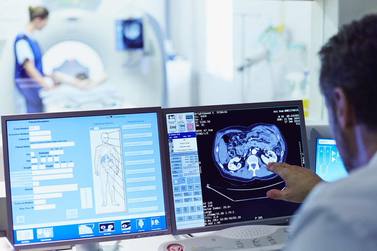
Computed tomography, often known as CT scanning, creates cross-sectional images of the body using X-rays and powerful computer technology. Newer CT methods, such as dual-energy CT, enable clinicians to distinguish between tissues based on their chemical makeup. Some clinics now offer spectral CT scanning, which allows for more detailed material decomposition than dual-energy CT.
Cone-beam CT is another breakthrough that creates high-resolution 3D images of small body parts such as the head, neck, and extremities. This technique is ideal for dentistry and orthopedic imaging applications. Some clinics have also implemented CT equipment that can perform CT-guided treatments, allowing for minimally invasive interventions guided by images.
Interpreting advanced imaging findings
While new technologies produce more detailed scans, correctly analyzing these complex images takes specialist knowledge. The Best diagnostic imaging clinic invests in cutting-edge equipment and hires highly trained radiologists with subspecialty certification in a variety of modalities. Many radiologists pursue extra fellowship training in specific fields such as neuroradiology, body imaging, nuclear medicine, and more.
Some clinics employ radiologists who specialize in quantitative imaging, which uses modern computer techniques to extract more information from scans. Some clinics are also starting to use artificial intelligence and deep learning to help radiologists discover minor abnormalities while lowering diagnostic errors and missed results.
Multi-modality Imaging Centers
Some clinics have elevated diagnostic imaging to a new level by combining numerous modalities under one roof. These multi-modality centers house cutting-edge equipment for MRI, CT, PET/CT, mammography, ultrasound, and other technologies in a single, central location.
Coordinated Care via Imaging
Having all modalities nearby improves the patient experience and enables for more coordinated care. Patients frequently require many types of scans on the same day. Patients at multi-modality centers can conveniently undergo all essential exams without having to travel between locations.
Specialization within Specialization.
Given the wide range of imaging modalities, sub-specialization is becoming increasingly significant. Important multimodality centers can accommodate teams of sub-specialized radiologists focused on specific topics. Some companies, for example, hire breast imaging radiologists who specialize in mammography and breast ultrasound only.
Others employ cardiothoracic imaging radiologists, body MRI radiologists, or neuro interventional radiologists to do minimally invasive operations. This high level of specialization assures that only the most competent interpreters analyze each patient’s scans.
Research and Development Capabilities
Top diagnostic imaging clinics contribute to the continual development and evaluation of new technologies. Some multimodality centers have on-site research laboratories. Radiologists and engineers work together to evaluate prototype equipment and imaging techniques.
These facilities also host valuable clinical trials. For example, a new MRI contrast agent could be assessed via a trial scan of healthy participants. The results aid in the optimization of the contrast agent before its application in patients. The findings of the study will lead to improved care in the future.
Setting New Standards
Multi-modality imaging centers are genuinely at the forefront of radiology because they combine the whole range of modern diagnostic equipment, specialist expertise, coordinated treatment, and research and development capabilities. Their integrated strategy establishes the gold standard for integrating cutting-edge technologies to improve diagnostic accuracy and patient outcomes. As imaging advances, these comprehensive centers will remain at the cutting edge.
Conclusion
Diagnostic imaging is rapidly evolving, pushing the boundaries of what the human eye can see. The most technologically equipped clinics in the United States combine cutting-edge equipment from several suppliers with specialized, skilled radiology staff. This combination of cutting-edge hardware and highly qualified personnel improves diagnosis accuracy, which benefits patient care. While technology is vital, human knowledge is still required to effectively utilize new imaging capabilities and achieve the best therapeutic outcomes.